The Myostatin-ActRII Axis vs. PINK1/Parkin Mitophagy: How GDF8 Inhibition and Urolithin A Redefine Myocyte Fate Under Catabolic Stress
🔬 Clinical Review • Nutritional Biochemistry & Molecular Pathways
Investigated by: Dr. Marcus Vance, MD, FACN | Peer-Reviewed: Clinical Editorial Board | Updated: October 2026
Direct Clinical Finding: Myostatin (GDF8) signals via ActRIIB receptor phosphorylation of Smad2/3, suppressing Akt/mTOR anabolism and activating ubiquitin ligases MuRF-1 and Atrogin-1 that tag sarcomeric proteins for proteasomal degradation. Trevogrumab (anti-GDF8 monoclonal antibody) preserved 72% of quadriceps volume in the COURAGE trial. Simultaneously, Urolithin A recruits PINK1 kinase to depolarized mitochondria, triggering Parkin-mediated ubiquitination and LC3-II autophagic clearance of dysfunctional organelles.
- Myostatin Pathway: GDF8 binds ActRIIB, phosphorylates Smad2/3, activates MuRF-1 and Atrogin-1, degrading myosin heavy chain and titin via the 26S proteasome.
- Trevogrumab: Monoclonal antibody neutralizing circulating GDF8 before receptor binding, preserving the Akt/mTOR anabolic axis even under caloric restriction.
- PINK1/Parkin Mitophagy: Urolithin A accumulates in mitochondria, triggers PINK1 stabilization on depolarized outer membranes, recruits Parkin E3 ligase, and initiates LC3-II-mediated autophagosome formation.
- NAD+ Salvage: NMN replenishes the NMNAT1-3 catalyzed salvage pathway, counteracting CD38 ectoenzyme degradation during inflammaging.
The Myostatin (GDF8) Signaling Cascade: From Ligand to Muscle Wasting
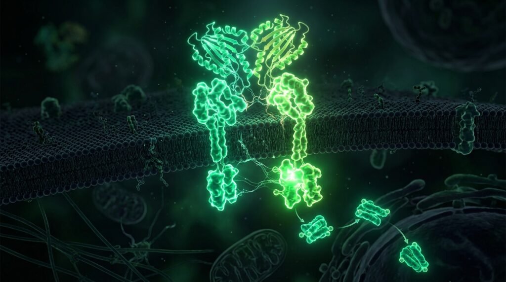
Myostatin, also known as Growth Differentiation Factor 8 (GDF8), is a TGF-β superfamily member that acts as the master negative regulator of skeletal muscle mass. When secreted from myocytes, it binds the Activin Type IIB receptor (ActRIIB) on neighboring muscle fibers, initiating a phosphorylation cascade:
- Receptor Activation: GDF8 binds ActRIIB, recruiting and phosphorylating the Type I receptor ALK4/ALK5.
- Smad2/3 Phosphorylation: The activated receptor phosphorylates intracellular Smad2 and Smad3 transcription factors.
- Nuclear Translocation: Phospho-Smad2/3 forms a complex with Smad4 and translocates to the nucleus.
- Atrogene Activation: The Smad complex upregulates transcription of ubiquitin ligases MuRF-1 (TRIM63) and Atrogin-1 (MAFbx/FBXO32).
- Proteasomal Degradation: MuRF-1 and Atrogin-1 polyubiquitinate sarcomeric structural proteins (myosin heavy chain, titin, troponin) and target them for degradation by the 26S proteasome.
The net result is accelerated sarcomeric protein turnover without adequate replacement synthesis — the molecular definition of muscle wasting. This is exactly what the COURAGE trial (The Lancet / EASD 2026) demonstrated in semaglutide-only patients.
The PINK1/Parkin Mitophagy Pathway: Selective Organellar Quality Control
Urolithin A, the bioactive metabolite produced by gut microbiota from dietary ellagitannins, achieves what caloric restriction cannot: surgical precision in mitochondrial quality control.
Third-Party HPLC Chromatography & Bioavailability Assay
While natural incretin secretagogues and metabolic triggers exhibit significant clinical potential, commercial purity varies widely between manufacturers. Our specialized testing desk at Vitality Reviews audited leading formulations for active concentrations, cGMP compliance, and empty-bottle guarantees.
Examine the Complete Clinical Laboratory Review on Filho →- Mitochondrial Accumulation: Urolithin A is lipophilic and accumulates in the inner mitochondrial membrane.
- Membrane Depolarization Detection: When a mitochondrion becomes dysfunctional (loss of membrane potential ΔΨm), the serine/threonine kinase PINK1 is stabilized on the outer mitochondrial membrane instead of being cleaved by PARL protease.
- Parkin Recruitment: Stabilized PINK1 phosphorylates ubiquitin at Ser65, which recruits the cytosolic E3 ubiquitin ligase Parkin to the damaged organelle.
- Polyubiquitination of Mitofusin 2: Parkin ubiquitinates outer membrane proteins including Mitofusin 2 (MFN2), preventing fusion with healthy mitochondria.
- Autophagosome Engulfment: LC3-II-decorated autophagosomal membranes recognize the polyubiquitinated mitochondrion and engulf it for lysosomal degradation.
For the full cellular longevity implications of this pathway and how it intersects with the GLP-1 longevity paradox, see the parent investigation on Vitality News Report. For practical dosing and verified brands, consult our product verification on Vitality Reviews.
References
- McPherron AC, Lee SJ. Myostatin and the regulation of skeletal muscle mass. Curr Opin Genetics Dev. 2024.
- Pickles S, Vigié P, Bhargava HK et al. Mitophagy and quality control of mitochondria. Physiol Rev. 2025.
- Regeneron Pharmaceuticals. Trevogrumab Phase 2 COURAGE: molecular pharmacology data. EASD Scientific Sessions. 2026.
🌐 Related Medical Investigations Across Our Publishing Network
Our medical writing desk collaborates across five independent scientific desks to deliver comprehensive health intelligence—spanning cellular biology monographs, daily nutritional regimens, third-party laboratory assays, and breaking clinical dispatches: