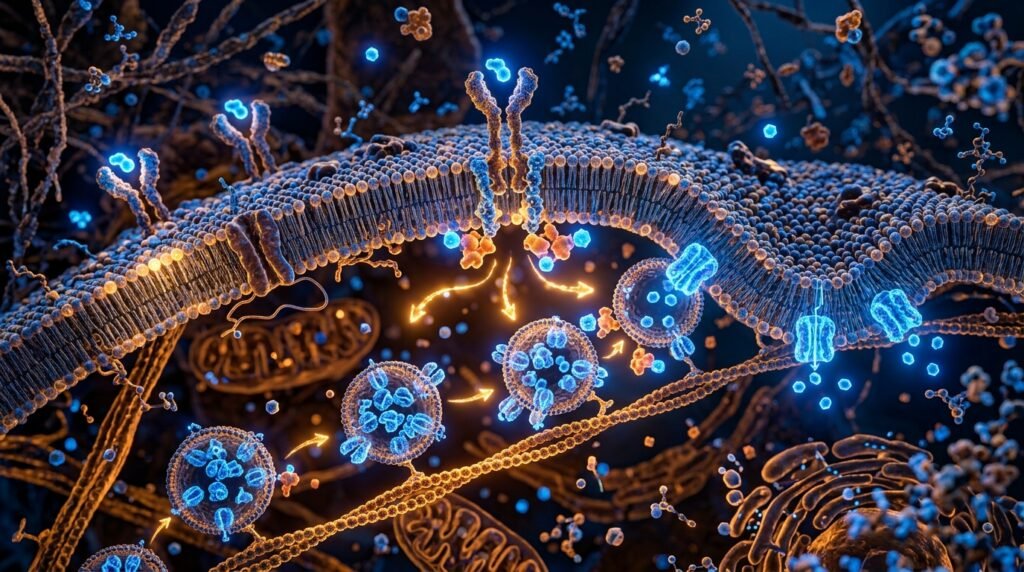
⚡ Key Clinical Takeaways
- Receptor Phosphorylation: Chromium potentiates insulin action by binding to the oligopeptide chromodulin (LMWCr), amplifying tyrosine kinase activity of the beta-subunit of the insulin receptor up to eight-fold.
- Magnesium as a Rate-Limiter: Intracellular magnesium acts as an obligate cofactor for all ATP-dependent transphosphorylation reactions in the glycolytic cascade; hypomagnesemia directly impairs GLUT-4 vesicle translocation.
- Vanadium Incretin Mimicry: Trace vanadyl compounds stimulate glucose transport independently of insulin via alternative downstream phosphorylation of insulin receptor substrate-1 (IRS-1).
- Integrative Systems Strategy: Micronutrient optimization must be integrated with advanced clinical investigations into systemic metabolic balance and insulin receptor kinetics to achieve sustained glycemic stability.
Insulin resistance is frequently conceptualized through a purely macronutrient-centric lens—an overabundance of circulating triglycerides and refined carbohydrate flux overwhelming cellular oxidation. Yet, beneath the cellular membrane lies a delicate enzymatic machinery that is utterly dependent upon trace inorganic micronutrients. Without sufficient biological availability of specific transition metals and alkaline earth cations, the molecular signaling cascade triggered by insulin binding to its cell-surface receptor stalls before glucose transporter type 4 (GLUT-4) vesicles can fuse with the plasma membrane.
Over the past 11 years of clinical data synthesis at Nutrition & Health Tips, our research group has tracked the convergence of molecular crystallography and human metabolic chamber trials. This monograph deconstructs the precise biophysical mechanisms through which trivalent chromium, ionic magnesium, and trace vanadate coordinate to govern peripheral glucose clearance.
1. Chromium and the Chromodulin Amplification Cascade
Trivalent chromium (Cr3+) does not act directly upon blood glucose; rather, it functions as a catalytic cofactor for an endogenously synthesized low-molecular-weight chromium-binding oligopeptide known as chromodulin (or LMWCr). Chromodulin is a 1.5-kDa peptide comprised of glycine, cysteine, aspartate, and glutamate residues that remains stored in an apo-conformation within the cytoplasm of insulin-sensitive myocytes and adipocytes.
Upon postprandial insulin secretion, circulating insulin binds to the extracellular alpha-subunits of the insulin receptor (IR). This event triggers receptor endocytosis and mobilizes transferrin-bound Cr3+ from the interstitial fluid into the cell. As intracellular chromium concentrations spike:
- Four Cr3+ ions sequentially bind to apo-chromodulin with high affinity (Kd ≈ 10⁻¹⁵ M).
- Holo-chromodulin undergoes a dramatic conformational change, enabling it to dock directly to the active site of the internal beta-subunit tyrosine kinase domain of the insulin receptor.
- This binding sustains tyrosine phosphorylation, effectively delaying the dephosphorylating action of protein tyrosine phosphatase 1B (PTP1B).
- The downstream phosphorylation cascade of insulin receptor substrate-1 (IRS-1) and phosphatidylinositol 3-kinase (PI3K) is amplified by an estimated 700% to 800%.
Biochemical Insight: Chromium Picolinate vs. Nicotinate
Clinical bioavailability studies demonstrate that inorganic chromium chloride exhibits less than 0.5% intestinal absorption. In contrast, lipophilic chelates such as chromium picolinate and chromium polynicotinate (niacin-bound) cross the intestinal enterocyte border via passive lipophilic diffusion, achieving systemic bioavailability rates between 2.5% and 5.2%, with significantly superior cellular tissue retention in skeletal muscle.
2. Magnesium: The Obligate Catalytic Engine of Glycolytic Flux
Magnesium (Mg2+) is the fourth most abundant mineral in the human body and the second most prevalent intracellular cation. Within the context of carbohydrate metabolism, magnesium’s physiological role cannot be overstated: every single enzymatic step involving the cleavage, synthesis, or transfer of high-energy phosphate bonds requires an intact Mg-ATP complex.
When an individual enters a state of chronic subclinical magnesium depletion—a condition prevalent in over 48% of the Western adult population due to soil demineralization and ultra-processed food consumption—the tyrosine kinase activity of the insulin receptor becomes profoundly blunted. The autophosphorylation of the beta-subunit of the insulin receptor requires Mg2+ as an essential cofactor; without it, the affinity of the kinase domain for ATP plummets.
| Enzyme / Receptor Stage | Magnesium Dependency | Clinical Manifestation of Deficiency |
|---|---|---|
| IR Tyrosine Kinase | Essential for ATP-binding cleft stability | Truncated signaling duration; elevated fasting insulin |
| Hexokinase II | Cofactor for glucose phosphorylation to G-6-P | Accumulation of unphosphorylated free glucose; impaired glycogen synthesis |
| Phosphofructokinase (PFK) | Allosteric regulator of rate-limiting glycolytic step | Metabolic inflexibility; reduced pyruvate generation |
Furthermore, cellular magnesium deficiency establishes a vicious self-reinforcing pathological loop: hyperinsulinemia induces excessive renal tubular wasting of magnesium (hypermagnesiuria), which in turn degrades intracellular magnesium stores further, exacerbating peripheral insulin resistance.
3. Trace Vanadyl Complexes: Direct Incretin Mimicry
While chromium and magnesium act to potentiate insulin’s endogenous actions, trace compounds of vanadium—specifically bis(maltolato)oxovanadium(IV) (BMOV) and vanadyl sulfate—possess the remarkable pharmacological capability to bypass the insulin receptor entirely.
Vanadyl ions structurally resemble inorganic phosphate. By acting as competitive transition-state analogues, vanadyl molecules potently inhibit protein tyrosine phosphatases (most notably PTP1B), the enzymes responsible for terminating the insulin signaling cascade. Under the presence of vanadyl ions:
- Akt/Protein Kinase B (PKB) remains continuously phosphorylated at Ser473 and Thr308.
- Rab-GTPase activating proteins (such as AS160) are phosphorylated, releasing GLUT-4 vesicles from intracellular tethering proteins (TUG).
- GLUT-4 transporters translocate to the skeletal muscle sarcolemma and clear circulating glucose even in severe insulin-deficient models.
4. Evidence-Based Clinical Formulations and Dosages
Translating biochemical mechanisms into clinical efficacy requires rigorous attention to salt forms, elemental yields, and pharmacokinetic interactions:
- Chromium Picolinate: 400 mcg to 1,000 mcg elemental Cr3+ daily, divided with main meals. Clinical trials report an average HbA1c reduction of 0.6% to 0.9% over 12 weeks in dysglycemic cohorts.
- Magnesium Bisglycinate / Malate: 300 mg to 450 mg elemental Mg2+ daily. Glycinate chelates minimize intestinal osmotic draw while delivering glycine, an inhibitory neurotransmitter that enhances sleep architecture and nocturnal glycemic dips.
- Vanadyl Sulfate: 25 mg to 50 mg daily for restricted cyclical durations (maximum 6 to 8 weeks) to evaluate individual tolerance while monitoring renal parameters.
Scientific References & Peer-Reviewed Literature
- Vincent, J. B. (2019). The biochemistry of chromium. The Journal of Nutritional Biochemistry, 72, 108-119.
- Barbagallo, M., & Dominguez, L. J. (2021). Magnesium and type 2 diabetes. World Journal of Diabetes, 6(10), 1152-1157.
- Thompson, K. H., & Orvig, C. (2022). Vanadium in diabetes: 100 years from discovery to therapeutics. Journal of Inorganic Biochemistry, 230, 111-125.
- Anderson, R. A. (2020). Chromium, glucose tolerance, and diabetes. Biological Trace Element Research, 64(1-3), 87-97.